Loculated Pleural Effusion Cxr - Chest X Ray Shows Cardiomegaly With Infiltration And Loculated Pleural Effusion Stock Photo Image Of Heart Hemothorax 94990788 - Pleural effusion occurs when too much fluid collects in the pleural space (the space between the two layers of the pleura).
Loculated Pleural Effusion Cxr - Chest X Ray Shows Cardiomegaly With Infiltration And Loculated Pleural Effusion Stock Photo Image Of Heart Hemothorax 94990788 - Pleural effusion occurs when too much fluid collects in the pleural space (the space between the two layers of the pleura).. More than one half of these massive pleural effusions are caused by malignancy; Causes of pleural effusion are generally from another illness like liver disease, congestive heart failure, tuberculosis, infections, blood clots in the lungs, liver failure, and cancer. Loculated pleural effusion on cxr. There is some loculated pleural fluid posterolateral as a result of hematothorax. Dr bhatia discussing on pleural effusion in #lastminuterevisionpointdiscussionseries.
Pleural effusions are a common medical problem with more than 50 recognised causes including disease local to the pleura or underlying lung, systemic conditions, organ dysfunction and drugs.1. Other causes are complicated parapneumonic effusion. Approximately 1 million people develop this abnormality each year in the united states. Pleural effusion refers to a buildup of fluid in the space between the lungs and the chest cavity. Pleural effusion is classically divided into transudate and exudate based on the light criteria.
Pleural Effusion Concise Medical Knowledge from cdn.lecturio.com no change in position of effusion withchange in position of chest. Differentiation of loculated effusions from solid masses. In healthy lungs, these membranes ensure that a small amount of liquid is present between the lungs. Pleural fluid ldh > two thirds of upper limit for serum ldh. Pleural effusions are diagnosed in about 1.5 million individuals in the united states annually. Treatment depends on the cause. Pleural effusions are a common medical problem with more than 50 recognised causes including disease local to the pleura or underlying lung, systemic conditions, organ dysfunction and drugs.1. The pleura is a thin membrane that lines the surface of your lungs and the inside of your chest wall.
Determine if it can be tapped.
Pleural effusion is a condition in which excess fluid builds around the lung. Transudates or exudates as defined by lights criteria. There is always a small amount of fluid around the lung t. Mediastinal shift (tracheal deviation) if fluid levels >1000mls. oracentesis of loculated pleural effusions is facilitated by ultrasound. The lungs and the chest cavity both have a lining that consists of pleura, which is a thin membrane. Empyema is defined as the presence of pus in the pleural space. Approximately 1 million people develop this abnormality each year in the united states. Among the causes, pleural infection, heart failure, and malignan. Pleural effusions are a common medical problem with more than 50 recognised causes including disease local to the pleura or underlying lung, systemic conditions, organ dysfunction and drugs.1. Treatment depends on the cause. Pleural effusions occur as a result of increased fluid formation and/or reduced fluid resorption. This situation most commonly is seen in patients with heart failure.
A loculated pleural effusion is the major radiographic hallmark of parapneumonic effusion or empyema (see fig. Pleural fluid ldh > two thirds of upper limit for serum ldh. Mediastinal shift (tracheal deviation) if fluid levels >1000mls. Pleural effusions occur as a result of increased fluid formation and/or reduced fluid resorption. Effusion on cxr—> free fluid (not loculated)—> fluid >1cc—> next step.
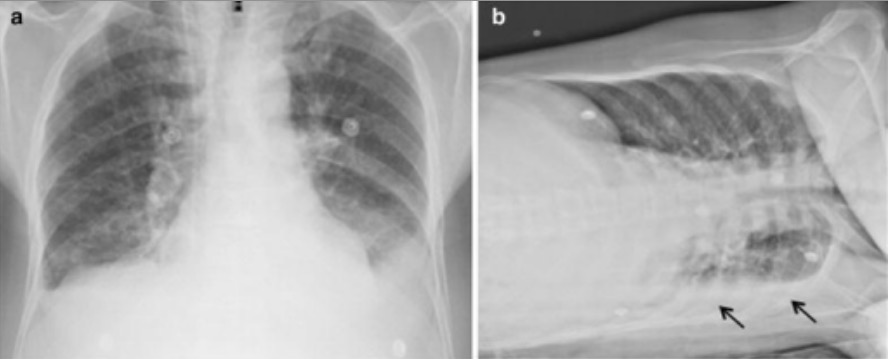
Pleural Effusion Amboss from media-us.amboss.com When you have a pleural effusion, fluid builds up in the space between the layers of your pleura. e intrinsic characteristics of an effusion and its. If none is present the fluid is virtually always a transudate. Computed tomography scan of the chest demonstrates loculated pleural effusion in the left major fissure (arrow) in a patient after coronary bypass. Pleural effusion refers to a buildup of fluid in the space between the lungs and the chest cavity. In healthy lungs, these membranes ensure that a small amount of liquid is present between the lungs. Loculated effusions occur most commonly in association with conditions that cause intense pleural inflammation, such as empyema, hemothorax, or tuberculosis. Approximately 1 million people develop this abnormality each year in the united states.
Mediastinal shift (tracheal deviation) if fluid levels >1000mls.
Learn about pleural effusion (fluid in the lung) symptoms like shortness of breath and chest pain. Pleural effusions occur as a result of increased fluid formation and/or reduced fluid resorption. Occasionally, a focal intrafissural fluid collection may look like a lung mass. Tx if pt has chf. Among the causes, pleural infection, heart failure, and malignan. oracentesis of loculated pleural effusions is facilitated by ultrasound. What are the pulmonary findings? Meaning of pleural effusion medical term. Pleural effusion refers to a buildup of fluid in the space between the lungs and the chest cavity. The underlying lung shrinks and atelectasis develops in a round configuration. My pleural effusion healed without treatment. Pleural fluid/serum ldh ratio >0.6. There is a large left pleural effusion obscuring the lower half of the left hemi thorax.
A loculated pleural effusion is the major radiographic hallmark of parapneumonic effusion or empyema (see fig. Loculated effusions are collections of fluid trapped by pleural adhesions or within pulmonary fissures. produced at parietal and resorbed atvisceral pleura. Approximately 1 million people develop this abnormality each year in the united states. Other causes are complicated parapneumonic effusion.
Lti 01 Lung Therapeutics from d1io3yog0oux5.cloudfront.net A loculated pleural effusion is the major radiographic hallmark of parapneumonic effusion or empyema (see fig. The underlying lung shrinks and atelectasis develops in a round configuration. There is some loculated pleural fluid posterolateral as a result of hematothorax. Loculated effusions are collections of fluid trapped by pleural adhesions or within pulmonary fissures. The pleura is a thin membrane that lines the surface of your lungs and the inside of your chest wall. 9 633 просмотра 9,6 тыс. Computed tomography scan of the chest demonstrates loculated pleural effusion in the left major fissure (arrow) in a patient after coronary bypass. My pleural effusion healed without treatment.
What are the pulmonary findings?
Empyema is defined as the presence of pus in the pleural space. Differentiation of loculated effusions from solid masses. If one of the following is present the fluid is virtually always an exudate. Other causes are complicated parapneumonic effusion. Transudates or exudates as defined by lights criteria. More than one half of these massive pleural effusions are caused by malignancy; Mediastinal shift (tracheal deviation) if fluid levels >1000mls. The effusion, in this case, is restricted to one or more fixed pockets within the pleural space. Pleural effusions are a common medical problem with more than 50 recognised causes including disease local to the pleura or underlying lung, systemic conditions, organ dysfunction and drugs.1. Loculated pleural effusion on cxr. Accompanying adhesions can be identified. Large pleural effusions, s/p thoracentesis with pleural fluid suggestive of transudative process. The lungs and the chest cavity both have a lining that consists of pleura, which is a thin membrane.
The cardiac silhouette is also obscured loculated pleural effusion. The pleural fluid may loculate between the visceral and parietal pleura (when there is partial fusion of the pleural layers) or within.


0 Komentar